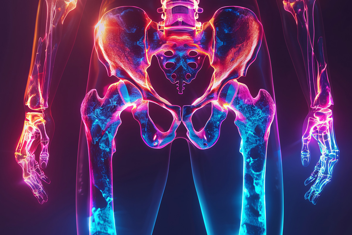
Magnetic resonance imaging of the thigh – a precise examination for diagnosing muscle injuries and pain

Magnetic resonance imaging (MRI) of the thigh is a safe and painless examination that provides detailed information about the muscles, tendons, nerves, blood vessels, and bones of the thigh. It is the best method for determining the exact cause of thigh pain, muscle injuries, tears, or stress-related problems.
The examination does not involve radiation, so it is also suitable for young people, athletes, and repeated monitoring.
When is magnetic resonance imaging of the thigh necessary?
Thigh pain and injuries can have many different causes, such as sudden sports injuries, strain, inflammation, or nerve compression. Magnetic resonance imaging (MRI) of the thigh provides a detailed view of tissue damage and possible changes that cannot be detected by other examinations.
Common reasons for MRI of the thigh:
- Sudden muscle strain or tear
- Recurring or prolonged thigh pain
- Suspected stress injury
- Swelling or hematoma of unknown cause
- Nerve compression or radiating pain
- Sports injury and assessment of its extent
- Accurate diagnosis prior to surgery or rehabilitation
Magnetic resonance imaging (MRI) of the thigh is particularly important for active individuals and athletes, as it helps assess the severity of an injury and speeds up a safe return to training.
How to prepare for a MRI scan of the thigh
Magnetic resonance imaging of the thigh does not require any special preparation. You can eat and drink normally before the examination. During the examination, the area must be free of metal objects and tight clothing.
If you have metal implants in your leg or elsewhere in your body, always report them in advance – the examination can usually be performed safely on the basis of an individual assessment.
How is magnetic resonance imaging of the thigh performed?
The examination is performed in a lying position. The leg to be imaged is positioned and supported so that it remains immobile during the examination. During magnetic imaging, the device makes a tapping sound, and you will be given earplugs or headphones.
The examination takes about 20–30 minutes. It is important to remain still so that the images are accurate.
Research results and follow-up measures
The images are analyzed by a radiologist who specializes in imaging examinations. You will receive a written report and the images within a few business days. They can also be sent directly to your doctor or physical therapist.
An MRI scan of the thigh helps to pinpoint the cause of the pain and guide you towards the right treatment, whether it is a sports injury, nerve compression, or prolonged pain.
You can easily book an appointment online – no referral is needed.
Visio Magneettikuvaus, Iso Omena, Espoo – Modern equipment and friendly customer service.
