MRI of the head and brain
Magnetic resonance imaging of the head and brain – a precise examination of the brain and nervous system
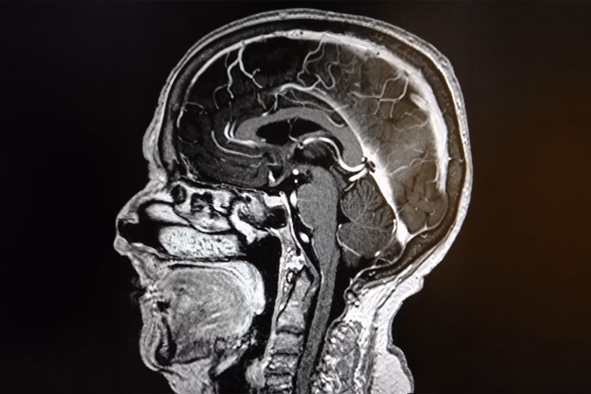
MRI of the head and brain is a safe, non-radiological examination that provides very precise information about the structure of the brain, brain chambers, cerebral blood vessels and skull. MRI is the preferred method for investigating the background of headaches, dizziness, memory problems or other neurological symptoms.
MRI of the brain uses a powerful magnetic field and radio frequencies to reveal tissue and nerve structures in detail – without the harmful effects of X-rays.
When is an MRI of the head and brain necessary?
Head and brain MRI is always used to rule out or detect neurological diseases such as brain tumours, inflammation, vascular abnormalities or early signs of memory disorders. It is also useful in identifying the cause of unexplained headaches, dizziness, balance problems or visual disturbances.
The most common reasons for MRI of the head and brain:
- Prolonged or abnormal headache
- Sudden dizziness or loss of balance
- Vision problems, double vision
- Memory problems or suspected memory loss
- Epileptic seizures or episodes of unconsciousness
- Post-accident assessment (e.g. concussion)
- Suspected brain haemorrhage or stroke
- How to prepare for an MRI scan of the head and brain
An MRI scan of the brain does not require any special preparation. You can eat and drink normally. It is important to remove all metal objects such as earrings, hairpins, glasses, prostheses and hearing aids when you come for the scan to ensure a safe and uninterrupted scan.
If you have a pacemaker, cochlear prosthesis or other implanted metal device, please let us know in advance. This will ensure that the examination can be carried out safely.
How is an MRI scan of the brain done?
The examination is performed in the supine position in an MRI machine with a mirrored imaging coil around the head. During the scan, the patient must remain still to obtain accurate images of the different structures of the brain.
The machine makes a tapping and humming sound during the examination, but you will be fitted with earplugs and headphones. The head and brain MRI scan takes about 20-30 minutes.
Results of the study and next steps
After the imaging, the radiologist, or radiologist, analyses the images and prepares a written report. You will usually receive the results within a few working days, and we can send them directly to your doctor if necessary.
An MRI scan of the brain can help identify any changes or diseases in time – and if necessary, point you in the right direction for further treatment.
Book an appointment for an MRI scan of your head and brain – without a doctor’s referral
Visio Magneettikuvaus, Iso Omena, Espoo – accessible location and friendly service.
You can book an appointment conveniently online, without a doctor’s referral.